The Whole Brain
This is a diagram of the whole brain. The main lobes are highlighted in different colors.
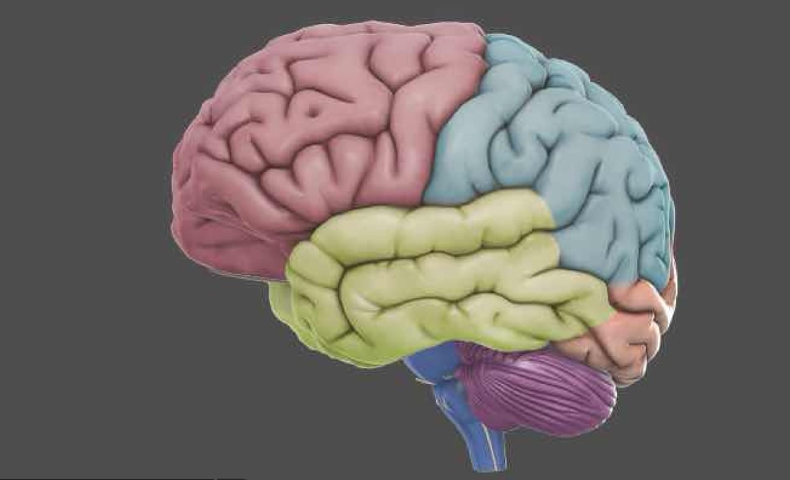

Temporal Lobe
Brain Stem
Cerebellum
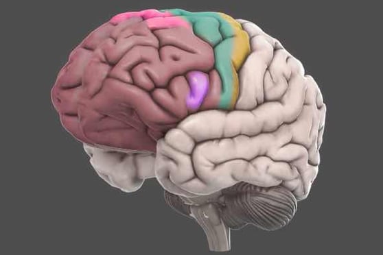

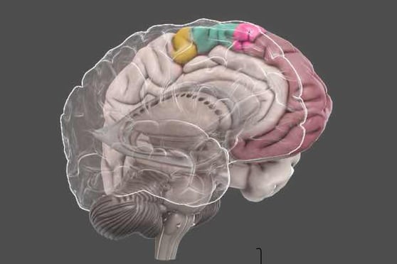

Frontal Lobe
The frontal lobes are part of the cerebral cortex and the largest of the brain's structures. They are the main site of 'higher' cognitive functions. The frontal lobes contain a number of important substructures, including the prefrontal cortex, orbitofrontal cortex, motor and premotor cortices and Broca's area. These substructures are involved in attention and thought, voluntary movement, decision making and language.
Although no longer a common practice, people have received a prefrontal labotomy in order to treat a variety of personality and cognitive disorders. This highly controversial practice involves destroying or severing the connections to the prefrontal cortex and often results in impaired voluntary behavior.
The Frontal Lobe focusses on executive processes (voluntary behavior such as decision making, planning, problem-solving and thinking), voluntary motor control, cognition, intelligence, attention, language processing, comprehension and many others.
The frontal lobes are the brain's largest structures and consequently have been associated with a large number of disorders. These include ADHD, schizophrenia and bipolar disorder (prefrontal cortex).
When damaged, they can cause paralysis, loss of spontaneity in social interactions, mood changes, inability to express language and atypical social skills / personality traits.


Temporal Lobes
The Temporal Lobes contain a number of structures which include functions such as perception, face/object recognition, memory acquisition, understanding language and emotional reactions. Functions associated with Temporal Lobes include recognition, perception (hearing, vision. smell), understanding language and learning/memory.
Damage to the temporal lobes can result in neurological deficits called agnosias, which refer to the inability to recognize specific categories (body parts, colors, faces, music, smells).
Interestingly, deep stimulation of the Temporal Lobes have been shown to produce profound religious and out-of-body experiences.
Schizophrenia is the cognitive disorder most closely aligned to temporal lobe dysfunction
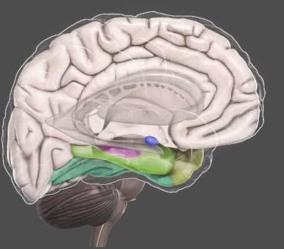



AMYGDALA
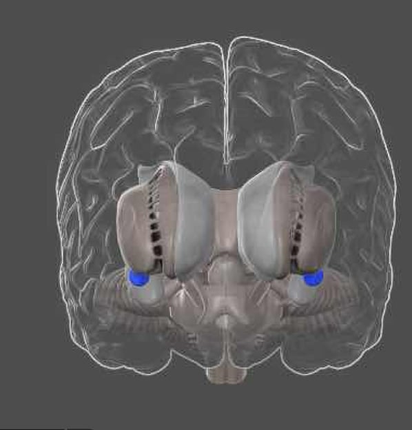

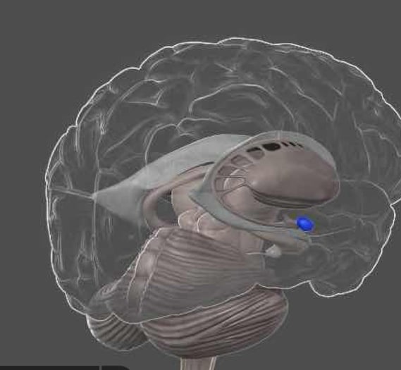
Amygdala from 3D Brain HQ
Amygdala from 3D Brain HQ
The amygdala is located next to the hippocampus, it is part of the temporal lobe and it's mainly involved in processing emotions. The amygdala is mainly involved with the fear response, and learning from fear. It links areas of the cortex that process higher cognitive information with hypothalamic and brain-stem systems that control 'lower' metabolic responses (e.g. touch, pain sensitivity and respiration). This allows the amygdala to coordinate physiological responses based on cognitive information- the most well known example is the 'fight-flight-freeze' response. Fear is the main emotion that the amygdala is known to control
Amygdala from 3D Brain HQ

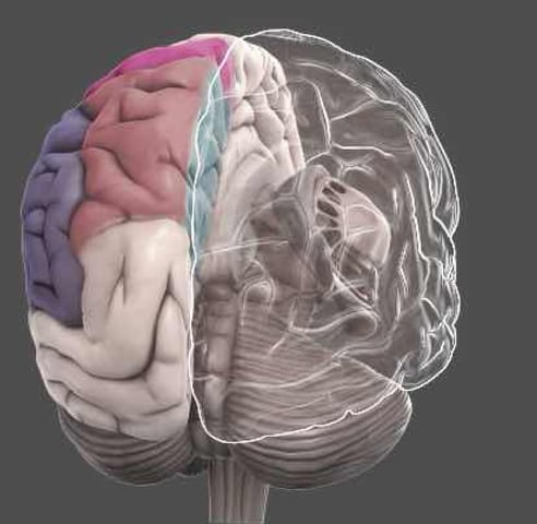

Parietal Lobes
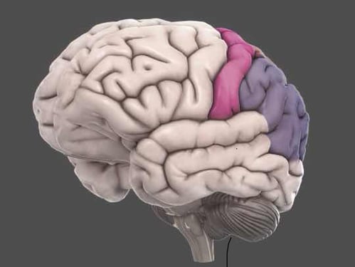
The parietal cortex plays an important role in integrating information from different senses to build a clear and detailed picture of the world. It brings together information from the ventral visual pathways (shows what things are) and the dorsal visual pathways (shows where things are located). This allows us to coordinate movements in response to the objects in our environment. It contains a number of distinct reference maps of the body, near space and distant space, which are constantly updated as we move and interact with the world.
The parietal cortex processes attentional awareness of the environment, is involved with manipulating objects and representing numbers.
Damage to the posterior parietal lobes can result in an intriguing neurological disorder called hemispatial neglect. The disorder is characterized by an inability to attend to people, objects, or one's own body on the side opposite the damaged area. Hemispatial neglect patients may eat from only one side of the plate, or dress one side of their body. In one case, (Bisiach and Luzzatti 1978), a report showing 2 patients who could not conjure visual images of buildings on the left side of their hometown- regardless of the direction they imagined themselves facing. This visual neglect, therefore, existed even in their imagination.
Associated functions include perception and integration of somatosensory (e.g. touch, pressure, temperature and pain); visuospatial processing; spatial attention; spatial mapping; number representation.
Associated cognitive disorders include atrophy in a number of brain structures, including the right temporo-parietal region, may be a precursor for Alzheimer's disease (Fouquet and colleagues, 2007). Vance and collegues (2007) found evidence of right parietal dysfunction in a subgroup of children with ADHD. Torrey (2007) reports an association between schizophrenia and the inferior parietal lobule.
When damaged, patients may not be able to locate and recognize objects/events or even parts of the body (hemispatial neglect). They may have difficulty discrimonating between sensory information; may become disorientated or have a lack of coordination.
Substructures include somatosensory cortex; inferior parietal lobule; superior parietal lobule; precuneus
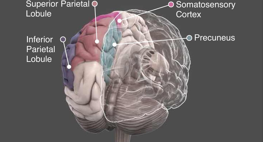

Superior Parietal Lobule
The superior parietal lobule is involved with spatial orientation, and receives a great deal of visual input as well as sensory input from one's hand. In addition to spatial cognition and visual perception, it has also been associated with reasoning, working memory, and attention.
Inferior Parietal Lobule
Inferior parietal lobule has been involved in the perception of emotions in facial stimuli, and interpretation of sensory information. The Inferior parietal lobule is concerned with language, mathematical operations, and body image, particularly the supramarginal gyrus and the angular gyrus.
Somatosensory Cortex
It is also known as Brodmann areas 1, 2, 3a, and 3b. Its primary function is to detect sensory information from the body regarding temperature, proprioception, touch, texture, and pain.
Precuneus
The precuneus is a brain region involved in a variety of complex functions, which include recollection and memory, integration of information (gestalt) relating to perception of the environment, cue reactivity, mental imagery strategies, episodic memory retrieval, and affective responses to pain.
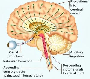
Reticular Activating System

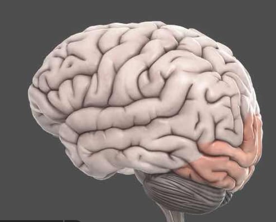

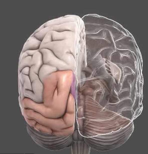

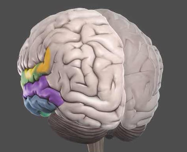
Occipital lobe from 3D Brain HQ
Occipital lobe from 3D Brain HQ
OCCIPITAL LOBE
The occipital cortex is the primary visual area of the brain. It receives projections from the retina (via the thalamus) from where different groups of neurons separately encode different visual information such as color, orientation and motion. Pathways from the occipital lobes reach the temporal and parietal lobes and are eventually processed consciously. The two important pathways of information originating in the occipital lobes are the dorsal and ventral streams.
Occipital lobe from 3D Brain HQ
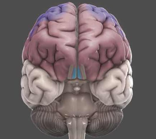

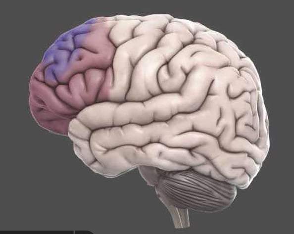

Prefrontal Cortex...
The prefrontal cortex is thought to play an important role in 'higher' brain functions. It is a crucial part of the executive system, which refers to planning, reasoning and judgement. It is also involved in personality and judgement by contributing to the assessment and control of appropriate social behaviors.
Prefrontal Cortex from 3D Brain HQ
Prefrontal Cortex from 3D Brain HQ
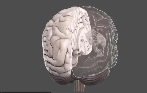

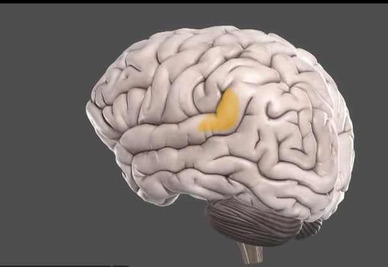

Wernicke's Area...
Wernicke's Area from 3D Brain HQ
Wernicke's Area from 3D Brain HQ
Wernicke's Area is a functionally defined structure that is involved with language comprehension. In most people, including a large majority of left-handed people, major language functions are contained in the left hemisphere of the brain. For most people, Wernicke's area is lateralized to the to the left side. It is named after Carl Wernicke, who worked with language-impaired patients to distinguish separate regions for language comprehension from production.
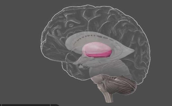

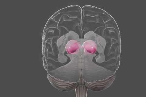

Thalamus...
The Thalamus is heavily involved in relaying information between the cortex, the brain stem and between different cortical structures. Because of this role in corticocortical interactions, the thalamus contributes to many processes in the brain- including perception, attention, timing and movement. It plays a central role in alertness and awareness.
Wernicke's Area from 3D Brain HQ
Wernicke's Area from 3D Brain HQ
Subscribe to hear when we post


